x
Quote information
Shipping address
Message
Your list is empty, add products to the list to send a request
Shop

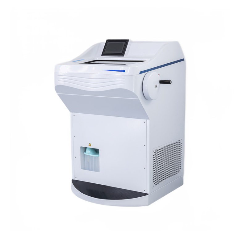
Cryostat Freezing Plus Microtome

See more by: Roundfin

A cryostat microtome is a precision instrument designed for the sectioning of frozen biological specimens in laboratory settings. It combines freezing technology with a microtome, allowing users to obtain thin slices of tissues for various applications in research, pathology, and diagnostics.

The Cryostat Microtome excels in preserving the structural integrity of delicate samples by maintaining low temperatures during the sectioning process. It is commonly utilized in histology and pathology laboratories for preparing tissue specimens for microscopic analysis. This specialized equipment is crucial for achieving high-quality, thin sections suitable for detailed examination under a microscope. Upgrade your laboratory capabilities with our advanced Cryostat Microtome, designed for efficient and precise frozen sectioning.

$0.00

10% off with your subscription Membership

Producer: Roundfin

Frequently bought together

Cryostat Freezing Plus Microtome

$0.00
10% off with your subscription Membership
Description

Specifications

Features

  • The specimen retraction function prevents specimens from accidental blade damage. This function can be turned on or off.
  • The unit can be defrosted manually or by using a controlled time setting.
  • A high-quality LED screen displays all user specification data.
  • The unit features high-brightness, low-power LED lights to provide stable and bright illumination.
  • It includes a UV sterilization function and is equipped with a cleaner to quickly collect wastes in the chamber.
  • The handwheel has 16 positions, ensuring safety.
  • The cryostat is equipped with two original German Danfoss brand compressors, providing special cooling for the chamber, freezing shelf, knife blade, and specifications.
  • A closed freezer cover plate ensures that cold in the cooling station is not lost.
  • The fully enclosed structure simplifies and makes cleaning up sample waste more convenient, giving a more refined appearance.
Key features include:
  • Fully enclosed design for the freezing chamber interior parts.
  • Frame cross-ball vertical section design with the transmission part placed in the freezer chamber. The vertical stroke can be 72mm, allowing a 22mm horizontal stroke for specimens, enabling cutting of larger samples.
  • The machine adopts a frame suspension structure, with the chassis shell serving only the role of protection and occlusion, ensuring stable operation during long-distance transport and precise slicing down to 1μm.
  • A large-size quick-freezing shelf allows the preparation of 26 specimens simultaneously, with two holes for semiconductor refrigeration.

Specimen retraction function: prevents specimens from accidental blade damage. This function can be turned off.

The unit can be defrosted manually or by a controlled time setting.

Introduction

A cryostat microtome is a specialized instrument used for cutting thin sections of frozen specimens, typically for histology and pathology research. The main parts of a cryostat microtome include:

  1. Freezing Chamber:

    • The compartment where specimens are frozen to a temperature low enough for sectioning.
  2. Microtome Sectioning Mechanism:

    • The cutting mechanism responsible for slicing thin sections from the frozen specimen. It includes a blade holder, advance/retract mechanism, and controls for adjusting section thickness.
  3. Specimen Holder:

    • A platform or clamp that holds the frozen specimen securely during the cutting process.
  4. Temperature Control System:

    • Components that regulate and maintain the low temperature inside the freezing chamber. This often includes a refrigeration system, temperature controls, and sensors.
  5. Knife Holder and Blade:

    • The knife holder supports the microtome blade, which is a sharp cutting tool that slices through the frozen specimen.
  6. Defrosting Mechanism:

    • A system for removing ice buildup or frost from the microtome components, allowing for smooth operation.
  7. Display and Controls:

    • User interface for setting and monitoring parameters such as temperature, cutting thickness, and other relevant settings.
  8. Illumination System:

    • Light source to illuminate the specimen during the sectioning process, aiding visibility.
  9. Waste Collection System:

    • A mechanism to collect and dispose of waste generated during the sectioning process.
  10. Vacuum System:

    • Some cryostat microtomes include a vacuum system to assist in holding the specimen in place and removing debris.
  11. Storage Compartments:

    • Spaces for storing additional blades, tools, and accessories.


Types of rotary Microtome

  1. Manual Cryostat Microtome:

    • Requires manual operation for setting cutting parameters, advancing the specimen, and other adjustments. It is suitable for users who prefer hands-on control over the sectioning process.
  2. Semi-Automatic Cryostat Microtome:

    • Combines manual control with some automated features, such as motorized advance/retract mechanisms or automated temperature control. This type offers a balance between manual flexibility and automated convenience.
  3. Fully Automatic Cryostat Microtome:

    • Provides a high level of automation for various cutting parameters, including section thickness and specimen advance. This type is suitable for high-throughput applications where consistent and precise sectioning is crucial.
  4. Freezing Microtome:

    • Specialized for cutting sections from frozen specimens. It includes a freezing chamber to maintain low temperatures during sectioning.
  5. Infrared Cryostat Microtome:

    • Uses infrared technology to assist in the rapid freezing of specimens, providing efficient and precise sectioning capabilities.
  6. Digital Cryostat Microtome:

    • Integrates advanced digital controls and interfaces for setting and monitoring cutting parameters. It often includes digital displays and programmable settings for improved precision.
  7. Cryostat Microtome with UV Disinfection:

    • Equipped with a UV sterilization function to disinfect the cutting area and minimize the risk of contamination during sectioning.
  8. Clinical Cryostat Microtome:

    • Specifically designed for clinical pathology applications, offering features tailored to the needs of diagnostic laboratories.
  9. Research Cryostat Microtome:

    • Geared towards research laboratories, these models may have advanced features and capabilities suitable for a wide range of scientific investigations. 
Strengths and Limitations

Strengths:

  1. Precision Sectioning: One of the key strengths is its ability to provide high-precision sectioning of frozen tissue samples. This is crucial in histology and pathology applications where thin and accurate sections are required for analysis.

  2. Cryogenic Cooling: The incorporation of cryogenic cooling ensures that the samples remain frozen during the sectioning process. This is particularly advantageous for preserving the integrity of delicate structures within the tissue.

  3. Versatility: Cryostat Rotary Freezing Microtomes are versatile instruments that can handle a variety of sample types, including soft and hard tissues. This makes them suitable for a wide range of research and diagnostic applications.

  4. Speed: Compared to traditional microtomes, cryostat rotary microtomes can provide quicker results due to the rapid freezing and sectioning process.

  5. Reduced Artifacts: The frozen sectioning process helps reduce artifacts that may be introduced by other methods, allowing for more accurate analysis of tissue structures.

Limitations:

  1. Cost: Cryostat Rotary Freezing Microtomes can be relatively expensive to purchase and maintain, making them less accessible for some research labs or smaller institutions with budget constraints.

  2. Complex Operation: Operating a cryostat microtome may require specialized training due to its complex design and the need to manage cryogenic temperatures. This can limit its use to trained personnel.

  3. Sample Size Restrictions: The size of the samples that can be accommodated may be limited by the dimensions of the cryostat chamber. Larger specimens may need to be processed differently.

  4. Maintenance Requirements: Cryostat microtomes require regular maintenance, including defrosting and cleaning, to ensure optimal performance. Failure to maintain the instrument properly can lead to issues with sectioning quality.

Summary
  • Specimen retraction function for preventing accidental blade damage, with the option to turn it on or off.
  • Manual or controlled time setting for defrosting the unit.
  • High-quality LED screen displaying user specification data.
  • UV sterilization function and waste collection cleaner for chamber hygiene.
  • 16-position handwheel for enhanced safety during operation.
  • Equipped with two original German Danfoss compressors for specialized cooling of various components.
  • Fully enclosed design for the freezing chamber interior, simplifying cleaning and providing a refined appearance.
  • Frame cross-ball vertical section design with a large stroke for cutting larger samples.
  • Frame suspension structure ensures stable operation during transport, achieving precise slicing down to 1μm.
  • Quick-freezing shelf for simultaneous preparation of 26 specimens, featuring semiconductor refrigeration.
  • Various types of cryostat microtomes available, including manual, semi-automatic, fully automatic, freezing, infrared, digital, and those with UV disinfection.
  • Clinical and research-specific cryostat microtomes catering to different laboratory needs.
References
  1. Tosney, K. W., & Landmesser, L. T. (1986). Neurites and growth cones in the chick embryo. Enhanced tissue preservation and visualization of HRP-labeled subpopulations in serial 25-microns plastic sections cut on a rotary microtomeJournal of Histochemistry & Cytochemistry34(7), 953–957. https://doi.org/10.1177/34.7.3519758 
  1. Sharma, R. (2022). Microtomy. Retrieved February 5, 2022, from Slideshare.net website: https://www.slideshare.net/candysharma777/microtomy-67236029
  2. Frimiano, E M S, N, C. N., A, V. D., Sales, A., Mendes, & A, N. A. (2022). Histological study of the liver of the lizard Tropidurus torquatusWied 1820, (Squamata: Tropiduridae)(AU). Braz. J. Morphol. Sci, 165–170. Retrieved from https://pesquisa.bvsalud.org/portal/resource/pt/lil-644137
  3. Muhd-Farouk, H., Amin-Safwan, A., Mahsol, H., & Ikhwanuddin, M. (2017). Histological Characteristics on the Testes of Mud Spiny Lobster, Panulirus polyphagus (Herbst, 1793). Pakistan Journal of Biological Sciences: PJBS20(7), 365-371. https://doi.org/10.3923/pjbs.2017.365.371
  4. Sy, J., & Ang, L.-C. (2018). Microtomy: Cutting Formalin-Fixed, Paraffin-Embedded SectionsMethods in Molecular Biology, 269–278. https://doi.org/10.1007/978-1-4939-8935-5_23

Additional information

Brand

Roundfin

Net weight

150 kg

Size

800*700*1190 mm

Gross weight

175 kg

Packing size

860*920*1480 mm

Voltage and frequency

AC220V±10%,50Hz(Standard)
AC110V±10%,60Hz

Power

500W

Noise

50Db(A)

Refrigerany

R404A

Type for blade holder

Yes

Waste Cleaner

Yes

Handwheel lock

16 positions locked

Certification

CE

Other remarks

OEM/custom-made accepted

Reviews

There are no reviews yet.

Be the first to review “Cryostat Freezing Plus Microtome”